
If A Peacock’s Tail Flashes Electric Blue Then Goes Dark, Here’s The Nano-Grid Bending Light Instead Of Pigment
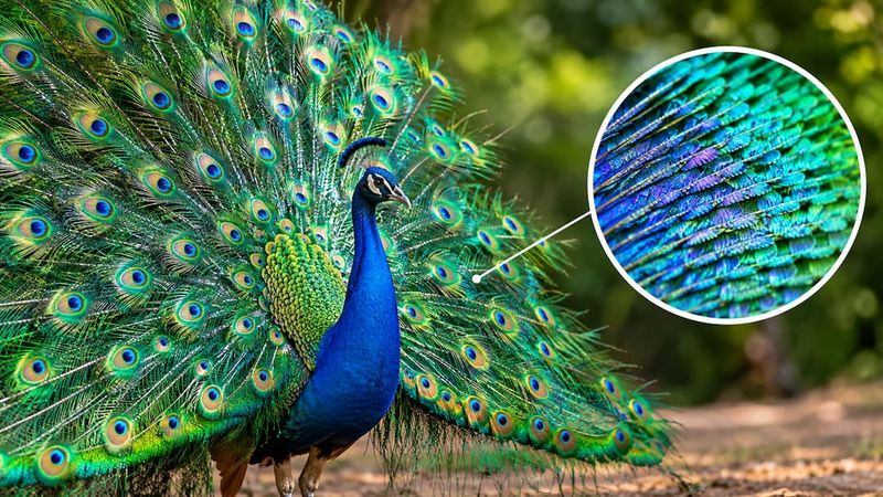
Watch a peacock fan its tail and you might swear the feathers are lit from inside, blazing electric blue one moment and fading to dull brown the next. No lamp is hidden in there, and no blue dye is doing the work either.
The secret is a microscopic architecture built from biological materials that selectively amplifies certain wavelengths of light, and the flash changes the moment you shift your angle. Understanding how that works reveals something surprising about color itself.
The flash changes with your position—not because the feather changes color instantly
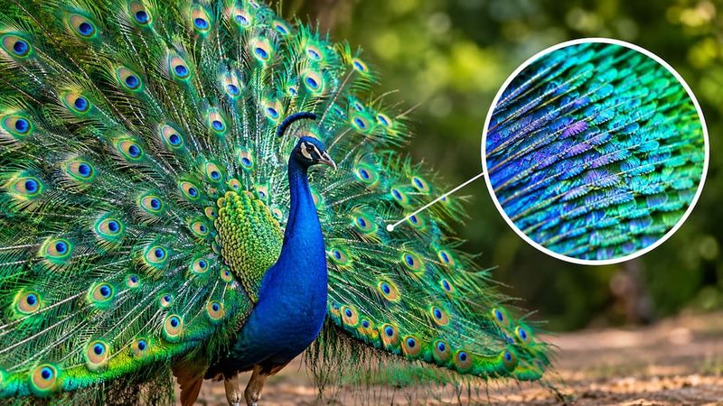
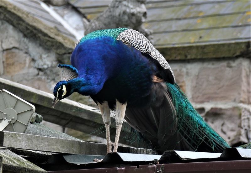
Stand directly in front of a displaying peacock and the tail blazes with blue-green light so intense it looks almost electric. Step a few feet to the side and the same feathers can look olive, bronze, or unremarkable brown.
The feather did not change. Your position relative to the feather and the light source did, and that shift altered which wavelengths of reflected light reached your eyes.
Peacock iridescence is primarily structural rather than produced by a conventional blue-green dye. The vivid color comes from a nanoscale architecture inside the feather’s barbules, the fine hairlike branches that extend from the main feather shaft.
That architecture reflects and reinforces particular wavelengths of light with remarkable precision, but only when the geometry of the feather, the observer, and the light source lines up correctly.
Research on iridescent neck and breast feathers of Pavo cristatus confirmed that the reflected hue and brightness shift measurably as the observation angle changes. A separate study measuring angle-dependent iridescence across two peafowl species found that these changes are often gradual rather than all-or-nothing: the strongest flash may retreat from your line of sight while some color remains visible from a nearby angle.
Two common oversimplifications are worth correcting early. First, the color does not vanish the instant any angle changes; it can shift in hue, brighten, dim, or lose its most vivid peak depending on the precise geometry involved.
Second, calling the effect “pure geometry” is not quite accurate because biological pigments are physically part of the optical structure, not absent from it. The article that follows explains both the mechanism and those qualifications in detail.
The color-producing structure sits inside the barbules
Feather color produced by a surface coating would behave like paint: it would look the same from most angles and would not shift dramatically when you moved. Peacock iridescence behaves very differently, and the reason is that the color-producing machinery is embedded deep inside each barbule rather than spread across the feather’s outer surface.
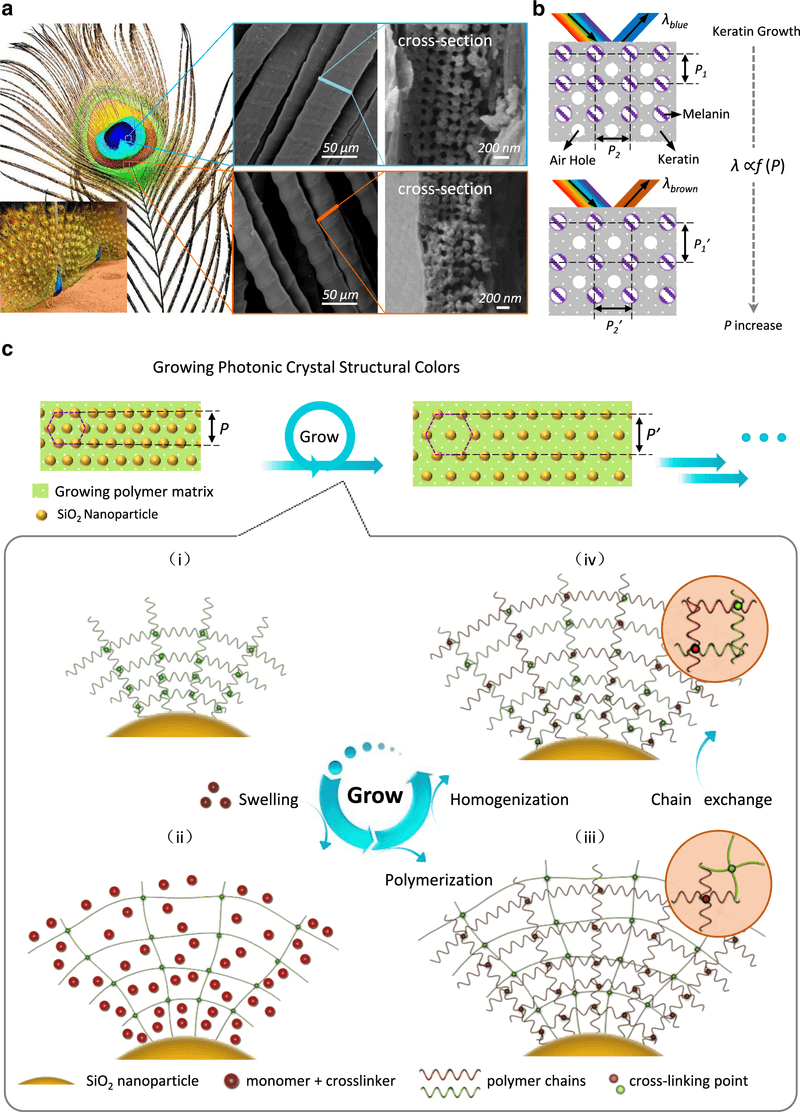
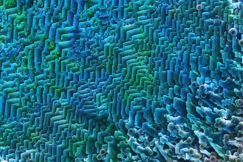
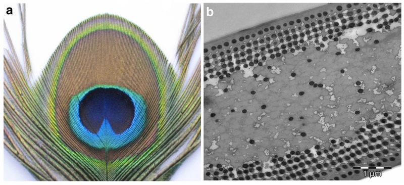
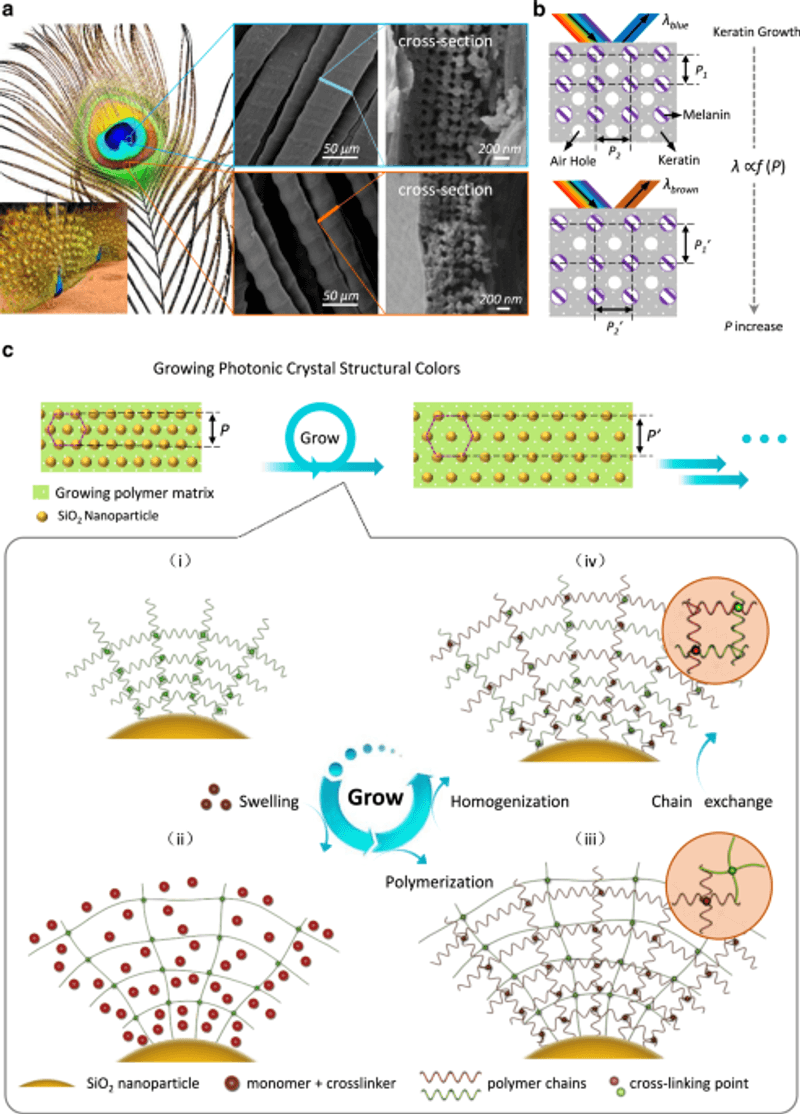
A barbule is one of the tiny, hair-thin branches that zip together to form the flat vane of a feather. Inside the cortex of each iridescent peacock barbule, a landmark study published in PNAS on coloration strategies in peacock feathers revealed a two-dimensional lattice: cylindrical melanin rods arranged in regular rows, surrounded by keratin and separated by air spaces.
The spacing of those rods, the thickness of the keratin layers between them, and the number of structural layers stacked on top of each other collectively determine which wavelengths of light the barbule reflects most strongly.
Keratin, melanin, and air each carry a different refractive index, meaning light travels through them at different speeds. When those materials alternate at precisely the right scale, they form what physicists call a photonic crystal, a periodic structure that interacts with light wavelength by wavelength rather than absorbing or scattering it uniformly.
Work comparing neck and breast barbule structures in Indian peafowl and a complementary Royal Society Interface study on peafowl iridescence both showed that differences in lattice period and layer count correspond to differences in the reflected color across feather regions. Wider spacing between melanin rods shifts the peak reflection toward longer, greener wavelengths; tighter spacing pushes it toward shorter, bluer ones.
The result is a color system governed by geometry at the nanometer scale, built from biological materials that are very much present inside every barbule.
How the lattice selects blue-green wavelengths
Visible light covers a continuous range of wavelengths, from violet at roughly 380 nanometers up through red at about 700 nanometers. A peacock barbule does not absorb all but one color the way a dye molecule does.
Instead, its periodic nanoscale lattice interacts with light through a combination of selective reflection, interference, and absorption that together amplify blue-green wavelengths and suppress others.
Picture the lattice as a stack of very thin, partially reflective layers. When a light wave enters the structure, a portion reflects off the first layer boundary, another portion travels deeper and reflects off the second boundary, and so on through many layers.
Whether those reflected portions add together or cancel each other depends on the wavelength of the light and the physical spacing of the layers. Wavelengths whose reflections arrive back in step with each other – a condition called constructive interference – are reinforced and emerge strongly.
Wavelengths that arrive back out of step cancel each other in destructive interference and are suppressed.
The lattice spacing inside peacock barbules is tuned so that blue and green wavelengths experience constructive interference across multiple layers, producing the vivid flash an observer sees. Wavelengths outside that range are either suppressed by destructive interference or absorbed by the melanin rods within the structure.
The PNAS coloration study measured the optical properties of barbule sections and showed that their reflectance spectra matched predictions from photonic-crystal models rather than from simple dye absorption curves.
Calling this mechanism “light bending” understates what is happening. A prism bends every wavelength by a slightly different amount and spreads them apart.
The photonic lattice in a peacock barbule instead selects specific wavelengths through interference across many periodic layers, reinforcing some and extinguishing others. The distinction matters because it explains why the reflected color is so pure and so sensitive to the precise geometry of the structure and the observer.
Why the flash shifts or fades at another angle
Constructive interference in a photonic crystal depends on the angle at which light enters the structure and the angle at which reflected light exits toward the observer. Change either angle and the effective path length through the lattice changes, which shifts the wavelengths that experience constructive interference.
The barbule’s internal architecture stays exactly where it is; what moves is the optical geometry connecting the light source, the feather, and the observer.
Measurements of angle-dependent iridescence across Indian and green peafowl showed that both hue and brightness shift as the observation angle changes, and the effect is not a simple on-off switch. From some positions the observer sees a strong blue-green peak; rotating slightly shifts that peak toward green or even yellow-green; moving further off the optimal angle reduces overall brightness while some color remains.
The flash fades from one line of sight without necessarily disappearing from all others simultaneously.
Several variables interact to produce what a specific observer actually sees at any moment. The illumination angle matters because it sets the direction light enters the barbule lattice.
The viewing angle matters because it determines which reflected wavelengths reach the observer’s eye. The feather’s own orientation in space matters because tilting the feather changes both simultaneously.
The region of the feather also matters because different parts of the eyespot have different lattice parameters and therefore different optimal angles, as discussed in a later section.
Background light and the spectrum of the light source add further complexity. Broad-spectrum white light gives the lattice more wavelengths to select from, producing a richer flash.
Narrow or colored illumination may shift or dull the appearance even at an otherwise ideal angle. Spectroscopic measurements of peacock barbules confirmed that the reflectance peak position and height both vary with illumination and observation geometry, reinforcing that angle dependence is a continuous, multivariable phenomenon rather than a simple color-on-or-off event.
A laboratory test separates structure from conventional dye
One of the clearest pieces of evidence that peacock barbule color is structural rather than produced by a conventional dye comes from a controlled optical experiment involving glycerin. The logic is straightforward: if a dye were responsible for the color, filling the feather’s air spaces with a liquid should not change the hue significantly, because the dye molecules would still absorb the same wavelengths.
If the nanoscale architecture is responsible, disrupting the refractive-index contrast between air, keratin, and melanin should measurably shift the reflected color.
Researchers in the PNAS coloration study infiltrated barbule samples with glycerin under controlled laboratory conditions. Glycerin has a refractive index much closer to keratin than air does, so filling the air spaces with it reduces the contrast between adjacent layers in the lattice.
The result was a measurable shift in the reflectance peak toward longer, redder wavelengths. The blue-green flash weakened or moved, precisely as photonic-crystal models predict when the refractive-index contrast is reduced.
A dye-only explanation cannot account for this result.
That finding is worth treating carefully in two respects. First, it is evidence, not proof of a single mechanism in isolation.
The experiment identified the air-space contrast as a critical optical variable, which strongly supports the structural explanation. Second, the glycerin infiltration was performed in a research setting with prepared barbule samples under controlled optical measurement conditions.
It is not a household activity and should not be treated as one. Soaking or wetting a feather with glycerin or any solvent at home would damage the sample, would not replicate the controlled optical geometry of the original experiment, and would tell an observer nothing reliable about the underlying mechanism.
The practical takeaway from the glycerin result is conceptual: the color depends on air being air, not on any colored molecule sitting inside the barbule. Remove the air contrast, and the photonic crystal’s tuning shifts.
That single experimental outcome separates peacock iridescence from conventional dye coloration more cleanly than almost any other test could.
Structural color still includes biological pigments
Structural coloration is sometimes described as though the feather contained no pigment at all, as if color were produced purely from transparent geometry. That framing is misleading in the case of peacock feathers.
Melanin rods are not incidental to the optical lattice; they are load-bearing members of it, and melanin is unambiguously a biological pigment.
Melanin absorbs light across a broad range of wavelengths, and that absorption does real optical work inside the barbule lattice. The PNAS coloration study and a related PubMed-indexed record of the same research program both describe melanin as a structural component whose absorptive properties help suppress unwanted wavelengths and increase the purity of the reflected color.
Without melanin’s absorption, stray wavelengths that were not fully cancelled by destructive interference would leak through and muddy the hue. The vivid, saturated quality of the blue-green flash depends in part on melanin doing its absorptive job alongside the lattice’s interference work.
A more recent layer of complexity comes from a 2026 study that identified lutein, a yellow carotenoid pigment, in iridescent peafowl feathers. That Animals journal research on structural coloration and carotenoids in peafowl proposed that lutein may act as a chemical filter, selectively absorbing wavelengths that would otherwise reduce the perceived saturation of the structural color.
If confirmed across additional specimens and conditions, this would mean that the final blue-green appearance results from at least three cooperating mechanisms: photonic-crystal interference, melanin absorption, and carotenoid filtering.
The safest summary of the current evidence is that nanoscale architecture supplies the primary optical mechanism for the vivid blue-green iridescence, while melanin is an inseparable structural and absorptive partner, and lutein may further refine the result. Calling the effect “pigment-free” is not accurate.
Calling it “primarily structural rather than produced by a conventional blue-green dye” captures what the evidence actually shows.
A peacock’s tail contains several optical systems
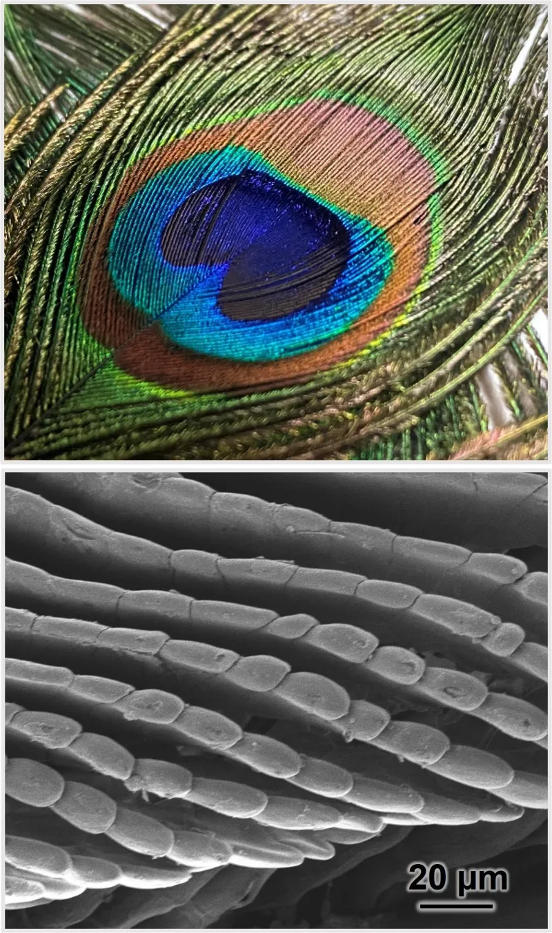
Saying “the peacock’s tail produces blue-green iridescence” is accurate as far as it goes, but the tail feather is not one uniform optical device. The famous eyespot alone contains multiple color zones – blue at the center, surrounded by rings of green, bronze, and brown – and each zone corresponds to a different set of structural parameters inside the barbules.
The PNAS coloration study showed that barbules sampled from different eyespot regions had measurably different lattice spacings and different numbers of structural layers. The blue center had a shorter lattice period tuned to shorter wavelengths, while the green ring had a longer period reflecting at greener wavelengths.
Brown regions presented yet another set of parameters, and a Physical Review E study on the structural origin of brown color in peacock barbules found that the brown regions use a partially disordered version of the lattice, producing broader, less saturated reflectance rather than the sharp iridescent peak of the blue-green zones.
Species distinctions matter as well. Most published structural-color research has focused on Indian peafowl (Pavo cristatus), the species familiar from parks and zoos worldwide.
Green peafowl (Pavo muticus) have a visually similar but structurally distinct feather architecture. Comparative Royal Society Interface work on peafowl iridescence and the two-species angle-dependent iridescence study both noted that the structural parameters and resulting color behavior differ between the two species.
Neck and breast feathers add still more variation, with their own barbule geometries producing their own reflectance profiles.
The practical implication is that the blue-green flash described throughout this article refers specifically to the iridescent zones of the Indian peafowl eyespot, which have been most thoroughly characterized. Claiming that every visible color on a peafowl tail shares a single identical mechanism would overstate what the research actually covers.
Watch the reflection move without altering the feather
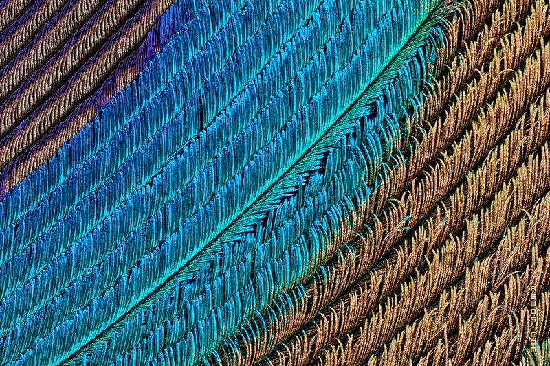
Measured angle-dependent reflectance in a laboratory is one thing; seeing it happen in your own hands is another. A legally obtained peacock feather under a single steady light source is all you need to observe the same phenomenon the researchers measured, with no chemicals, no cutting, and no special equipment required.
Set the feather on a table under a lamp that stays in one position. A bare incandescent or LED bulb works better than diffuse overhead fluorescent lighting because a point source creates a clear geometry between light, feather, and observer.
Keep the feather flat at first and look straight down at the iridescent zone. Then slowly tilt the feather toward you and away from you, watching the blue-green flash move across the surface.
Next, keep the feather still and shift your own head position side to side and up and down. Spectroscopic studies of peacock barbule reflectance and two-species angle-dependent measurements both found that the strongest flash occupies a relatively narrow angular range, so the brightest blue-green peak will appear and retreat as you move through different positions.
Keep the feather dry and intact throughout. Do not apply glycerin, water, oil, or any other liquid.
As described in an earlier section, the glycerin experiment was a controlled research procedure performed on prepared barbule samples; wetting a feather at home damages the structure and produces no useful optical information. Do not cut the barbules to examine the cross-section; the color depends on the intact periodic lattice, and cutting destroys the geometry you are trying to observe.
One important legal note before sourcing a feather: the U.S. Fish and Wildlife Service explains that possession of feathers from native North American birds is generally prohibited without authorization, and that restriction applies even to feathers found already molted on the ground.
Appearance alone cannot reliably establish a feather’s species or legal status. Peacock feathers from Indian peafowl, a non-native species in the United States, are widely sold legally, and purchasing from a reputable supplier is the straightforward way to obtain a feather with no legal ambiguity.
The structural architecture documented in the PNAS study is fully present in commercially available feathers, so a purchased feather will show the same angle-dependent flash as any other.
The structure stays put while the viewing geometry changes
After covering the mechanism from several angles, the core answer comes down to one precise statement: the nanoscale lattice inside each barbule is physically fixed in place. What changes is the geometric relationship between the light source, the feather, and the observer, and that change determines which wavelengths of reflected light travel toward any given pair of eyes.
The photonic-crystal architecture documented in peacock barbules reflects and reinforces specific wavelengths through constructive interference, while melanin absorption within the same structure suppresses stray wavelengths and sharpens the perceived color. The flash does not literally disappear when the angle changes; it redirects.
From a different position, an observer intercepts a different portion of the reflected light, which may carry a shifted hue, reduced brightness, or no strong peak at all from that particular line of sight. The color is still being produced in the feather, just not in the direction that observer is standing.
Angle-resolved measurements across two peafowl species confirmed that this redirection is gradual and continuous rather than a sudden switch. Meanwhile, the 2026 report on carotenoids in peafowl feathers adds that lutein may further shape which wavelengths emerge most strongly, suggesting the final color is a collaboration between the periodic lattice, melanin’s absorptive role, and possible carotenoid filtering.
Calling the peacock’s blue-green flash purely geometric, or purely pigment-free, misses the cooperation among all three. What the feather actually contains is a finely tuned optical system whose most dramatic property is that it sends its most vivid signal only to observers standing in exactly the right place.