
If Your Beehive Neighbors Have You Curious, Here’s The Precise Way Bee Venom Locks Onto Cancer Cells
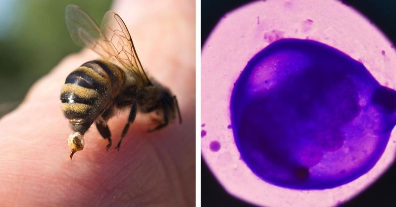
Honeybees are fascinating neighbors, and lately their venom has been showing up in cancer headlines that sound almost too good to be true. Scientists have found that melittin, the main toxic ingredient in a bee’s sting, can damage cancer cells in laboratory dishes and animal experiments.
But the story is more complicated than “bee venom targets cancer,” and understanding the difference could matter a great deal to anyone curious about what the research actually shows.
The precise targeting claim is the part researchers still need to prove
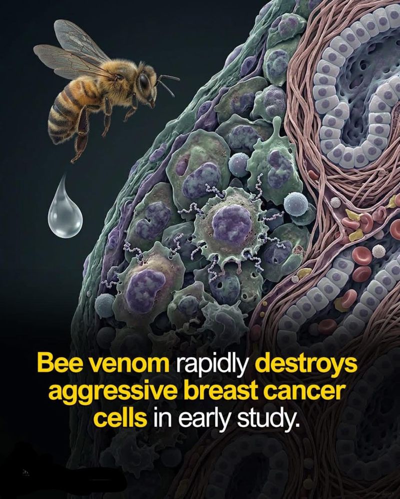
Headlines love the word “precise,” but in the case of bee venom and cancer, precision is the research goal, not the current reality. Melittin, the major toxic peptide in honeybee venom, is a short chain of 26 amino acids that carries a strong positive charge and can insert itself into the fatty layer surrounding nearly any cell.
That physical property is what makes it so potent – and so indiscriminate.
Researchers have observed that melittin can damage cancer cells in culture dishes and reduce tumor growth in some animal models. What those studies do not show is that native bee venom or free melittin automatically homes in on cancer cells while skipping healthy ones.
The mechanism is broad membrane disruption, not a guided search for malignant tissue.
Reviews of melittin’s cancer-related activity consistently describe what researchers call differential toxicity: some cancer cells appear more vulnerable than certain normal-cell comparators under specific laboratory conditions. That relative vulnerability is real and scientifically interesting.
It is not the same as a molecular address that tells melittin to attack tumors and leave everything else alone. Normal cells and red blood cells can be damaged too, which is a central problem the field is still working to solve.
The honest framing is that researchers are investigating whether melittin can be engineered into something tumor-selective. The bee’s sting, as it exists in nature, is not that thing yet.
How melittin breaches a cell membrane
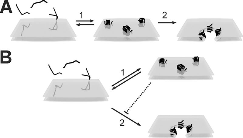
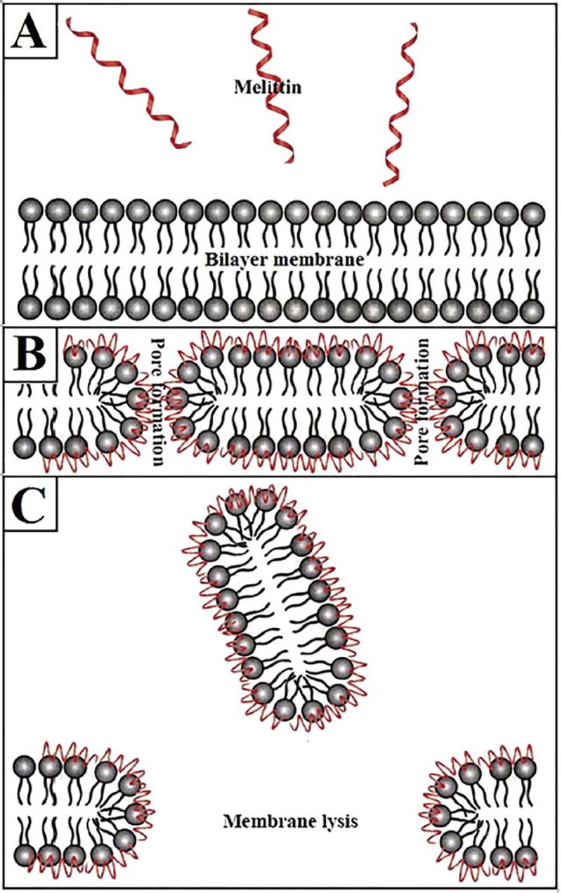
Picture a cell membrane as two layers of fatty molecules arranged tail-to-tail, forming a flexible barrier around everything inside the cell. Melittin attacks that barrier in a remarkably systematic way, moving through several stages that scientists have mapped in careful detail.
The process begins with attraction. Melittin carries a net positive charge, and most cell membranes carry a slight negative charge on their outer surface, so the peptide is drawn toward the membrane electrostatically.
Once it arrives, it lies flat against the lipid surface and begins to change shape, shifting from a loose coil into a more rigid, rod-like structure. That shape change is what allows the next step.
After settling against the surface, melittin rotates and inserts one end of itself into the fatty interior of the bilayer. Studies of pore formation and translocation show that a small cluster of melittin molecules can then arrange themselves into a ring-like structure, creating a temporary channel – a pore – through which water, ions, and small molecules leak out of the cell.
At low concentrations, the cell may survive this disruption. As the peptide-to-lipid ratio rises, the pores grow larger and more numerous, and the membrane can begin to break apart entirely.
Thermodynamic studies of melittin binding to lipid bilayers confirm that aggregation and pore formation depend heavily on how much peptide is present relative to the amount of membrane available. Higher ratios produce more destructive outcomes, including full membrane disintegration, cell lysis, and death.
Understanding this sequence explains both why melittin attracts research attention and why it poses a toxicity problem. The membrane-disrupting action that can kill a cancer cell in a dish can just as readily damage a healthy cell or rupture a red blood cell.
The mechanism itself does not carry a preference for malignancy built into its chemistry.
Why some tumor membranes may be more vulnerable
If melittin attacks membranes without a built-in cancer detector, why do some laboratory experiments show it hitting cancer cells harder than normal ones? The answer lies in how cancer changes the surface of a cell, and why those changes might make certain tumors a slightly easier target.
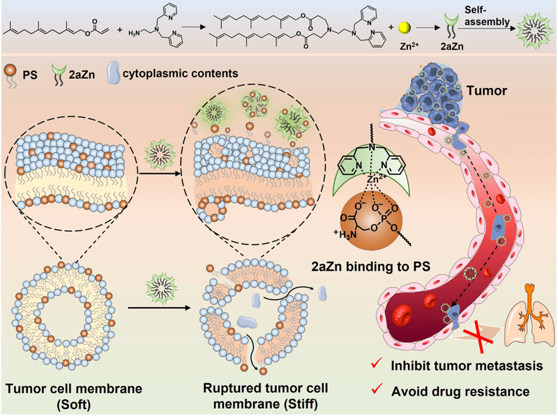
Healthy cells keep a lipid called phosphatidylserine tucked inside the inner leaflet of their membrane, essentially hidden from the outside world. Many cancer cells lose that organization and allow phosphatidylserine to flip to the outer surface.
Because phosphatidylserine carries a negative charge, its exposure increases the overall negative charge on the cancer cell’s outer face. Since melittin is positively charged, that shift can improve the peptide’s attraction to the cancer cell membrane compared with a normal cell that keeps phosphatidylserine properly tucked away.
Research into melittin binding thermodynamics shows that membrane composition, charge, and lipid organization all influence how readily the peptide inserts and forms pores. Cancer cells can also display altered cholesterol content and disordered lipid rafts, both of which may change membrane fluidity in ways that affect how easily melittin penetrates.
Some tumor cells also carry unusually dense coatings of glycosylated mucins, sugar-protein structures on their outer surface. Reviews of melittin’s application in cancer treatment suggest these mucin layers may interact with melittin in ways that draw it toward the cell rather than repelling it.
None of these features are universal. Not every cancer cell exposes phosphatidylserine, not every tumor has the same lipid organization, and some noncancerous cells share similar surface characteristics.
The advantage melittin gains from these membrane differences is real but probabilistic, more like a slight preference than a locked recognition system. Calling it a cancer-specific address overstates what the surface chemistry actually provides.
What the breast-cancer experiments actually showed
One of the most-cited recent studies on melittin and cancer involved breast-cancer cell lines, and its findings are genuinely striking – as long as they are read carefully and not stretched beyond what was tested.
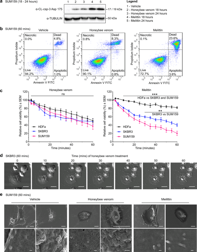
The 2020 breast-cancer study published in npj Precision Oncology tested both free melittin and crude honeybee venom against two aggressive breast-cancer cell lines – one HER2-enriched and one triple-negative – alongside a normal dermal fibroblast line as a healthy-cell comparator. Free melittin reduced the viability of the two cancer lines more rapidly than it affected the fibroblast line, and microscopy showed visible membrane disruption in the cancer cells within minutes of exposure.
That result is meaningful, but the study also found something that rarely makes the headlines: crude honeybee venom did not significantly distinguish between normal and cancer cells during the short exposure period tested. The whole venom, which contains dozens of compounds in addition to melittin, did not show the same preferential activity that the isolated peptide displayed.
The cancer lines also did not respond identically to each other, underscoring that even within a single cancer type, responses vary.
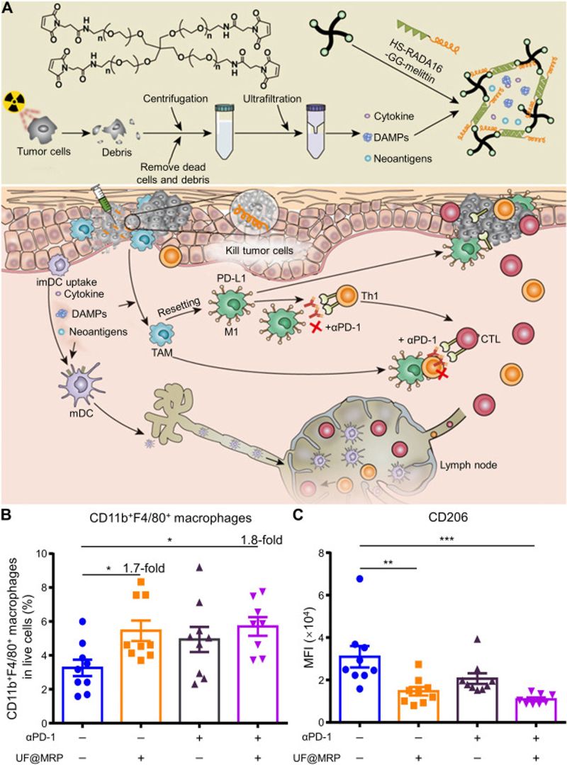
The researchers also ran mouse experiments in which melittin was injected directly into tumors alongside docetaxel, a standard chemotherapy drug. Combining both treatments produced better tumor control in the tested mouse model than either treatment alone.
That combination finding is biologically interesting and may point toward future research directions.
Every result in this study is preclinical. Cell-culture viability numbers and mouse tumor measurements are starting points for understanding a drug candidate, not evidence that a treatment works in human patients.
The study authors themselves framed their findings as a basis for further investigation, which is the appropriate scientific posture at this stage of research.
Where the apparent precision breaks down
The phrase “leaves healthy neighbors standing” is vivid, but the laboratory record does not support it as a description of how free melittin actually behaves. Variability in the results is the first sign that the targeting story is incomplete.
Responses to melittin shift depending on which cancer cell line is tested, which normal-cell line serves as the comparison, what concentration is used, how long the cells are exposed, and whether the peptide is free or packaged in a delivery system. The 2020 breast-cancer study found that some normal breast-cell models showed melittin sensitivity comparable to or even greater than certain cancer lines – a finding that directly contradicts the idea of automatic cancer preference.
The stronger differential appeared specifically when comparing aggressive triple-negative cancer cells against dermal fibroblasts, not across every pairing the researchers could have chosen.
A comprehensive review of melittin and its conjugates in cancer therapy lists nonspecific cytotoxicity, hemolysis, and systemic toxicity among the principal barriers to clinical use. Hemolysis – the rupturing of red blood cells – is particularly significant.
Red blood cells have membranes, and melittin disrupts membranes without checking whether the cell in question is cancerous. Systemic administration of free melittin would expose red blood cells throughout the body to that risk, which is one of the main reasons researchers cannot simply inject melittin into a patient’s bloodstream.
Beyond hemolysis, published analyses of melittin’s mechanisms document that the peptide can trigger apoptosis, necrosis, mitochondrial injury, and altered cell signaling in normal cells as well as cancer cells, depending on conditions. The concentration that damages a cancer cell in a dish may be close to the concentration that damages a healthy cell in the same dish.
That narrow window between “effective” and “harmful” is exactly the challenge delivery researchers are trying to solve, and it explains why the field has not already translated these findings into a usable therapy.
How researchers are trying to add a tumor address
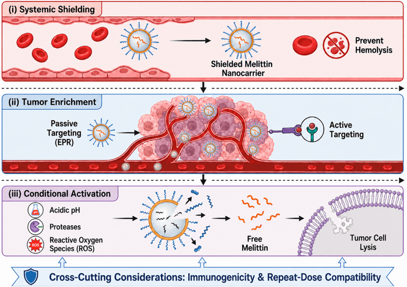
Because native melittin attacks membranes without a built-in cancer address, a growing field of research is focused on giving it one artificially. The engineering strategies vary widely, but they share a common goal: concentrate the peptide near tumor tissue and reduce its contact with everything else.
One of the most studied approaches uses lipid-coated nanoparticles as carriers. Research on molecularly targeted nanocarriers delivering melittin to tumor cells in mice showed that encasing the peptide inside a nanoparticle significantly reduced the nonspecific lysis that free melittin produces.
In mouse and laboratory models, these particles interacted preferentially with tumor or tumor-associated cells. The nanoparticle coating keeps melittin inactive during transit and releases it when the carrier reaches the target environment, a fundamentally different behavior from what happens when raw venom enters the body through a sting.
Other engineering approaches attach melittin to antibodies that recognize proteins found more abundantly on certain cancer cells. When the antibody binds its target on the tumor surface, it delivers melittin directly to that location.
Researchers have also explored masking strategies, where chemical groups block melittin’s membrane activity until enzymes present in the tumor microenvironment cleave the mask and reactivate the peptide.
Reviews of melittin conjugates in cancer therapy describe additional combinations including peptide-drug conjugates and melittin fused with tumor-homing peptide sequences such as RGD, which binds integrins overexpressed on some tumor blood vessels. The 2020 breast-cancer work also explored how combining melittin with standard chemotherapy might improve outcomes in animal models, pointing toward combination strategies as another research avenue.
Every one of these systems is an engineered drug candidate under laboratory or animal investigation. None of them is a bee sting, and none of them is crude honeybee venom.
The precision that may eventually emerge from this research would be a product of bioengineering, not a property that nature already built into the hive next door.
Why melittin is not yet a cancer treatment for people
Laboratory results and animal studies generate hypotheses; they do not generate treatments. The path from a promising cell-culture finding to a therapy that a doctor can prescribe is long, expensive, and full of failures, and melittin research has not completed that journey.
A 2024 review of bee venom composition and anticancer properties reported that no human clinical trials have established the effectiveness or cancer-treatment safety of bee venom or melittin. The evidence base remains predominantly in vitro (cell cultures) and in vivo in animal models, with unresolved challenges including peptide degradation in the bloodstream, systemic toxicity, hemolysis, immune reactions, allergenicity, and the difficulty of achieving effective concentrations at a tumor without harming surrounding tissue.
Reviews of melittin conjugates identify dosing and delivery as the field’s central unsolved problems. Even when engineered nanoparticle systems reduce off-target damage in mouse models, moving from a mouse experiment to a human trial requires demonstrating safety and efficacy across a far more complex biological system.
Many compounds that look impressive in mice do not survive that transition.
A separate confusion worth clearing up involves human bee-venom therapy studies. Some clinical research has examined bee-venom acupuncture or pharmacopuncture for pain management, inflammation, or supportive care in cancer patients.
Studies of bee-venom acupuncture address symptom management, not evidence that melittin shrinks or eliminates tumors. Conflating those two bodies of research overstates what either one actually shows.
The FDA is direct on this point: products claiming to cure cancer without regulatory approval can be dangerous and may delay care that has actually been proven to help. No reader should consider bee stings, injected venom, or commercially marketed bee-venom supplements as a substitute for oncology treatment.
The safe takeaway for anyone curious about bee venom
Melittin is genuinely interesting science. The peptide can exploit certain differences in tumor cell membranes, and researchers have documented real – if variable – differential activity against some cancer models.
That is worth knowing, and it is why serious laboratories continue to investigate it.
What melittin cannot do, at least not yet, is reliably distinguish every cancer cell from every healthy cell on its own. The precision that headlines describe is an engineering target being pursued through nanoparticles, antibody conjugates, and masking systems.
Targeted nanocarrier research in mice shows that engineered delivery can improve selectivity in animal models, but that is a laboratory result, not a clinical outcome. A bee sting delivers crude venom to skin and surrounding tissue; it does not perform precision oncology.
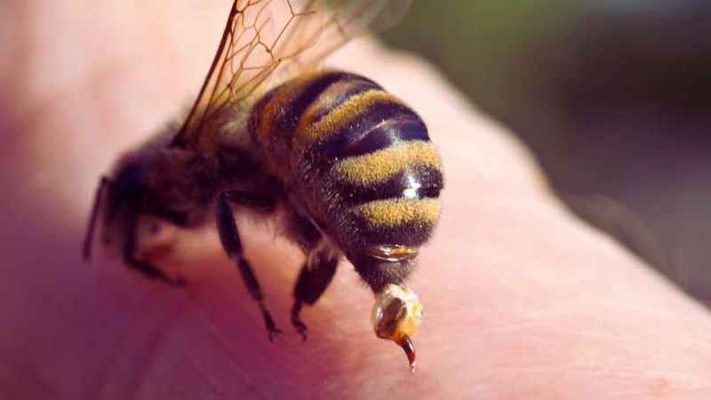
Anyone tempted to seek out bee stings, live-bee acupuncture, injected venom, or unapproved bee-venom supplements as a cancer remedy should know that the FDA warns these products can cause harm and delay proven treatment. Beyond the lack of cancer evidence, venom exposure carries real allergy risk.
Anaphylaxis – a severe, life-threatening allergic reaction – can occur even in people who have been stung before without incident.
If you or someone nearby shows signs of anaphylaxis after a sting, such as throat tightening, difficulty breathing, rapid heartbeat, or sudden dizziness, use an available epinephrine autoinjector immediately and call emergency services. The 2024 American Heart Association and American Red Cross first-aid guidelines recommend epinephrine as the first-line response, not antihistamines.
The bees next door may be fascinating neighbors, but their venom’s most reliable property is still the sting itself.